Recently, the research group led by Associate Professor Feng Jianjiang from the Department of Automation, Tsinghua University, collaborated closely with the team led by Professor Shi Heshui from the Department of Radiology of Wuhan Union Hospital of China, and successfully developed a CT-image-based AI identification and diagnosis system for COVID-19. It can accurately distinguish between COVID-19, influenza A/B pneumonia, non-viral pneumonia and non-pneumonia. The system achieved a 97.81% AUC value (area under curve) on a large-scale test set, and 92.99% and 93.25% AUC values on two independent domestic and overseas test sets.
The pneumonia caused by the novel coronavirus (Covid-19) has so far infected at least 40 million people worldwide and killed 1.11 million. To prevent and control the epidemic and treat patients in time, accurate and rapid diagnostic technology is critical. Chest CT imaging can directly display the status of pulmonary infection, which is the main imaging technique for COVID-19 diagnosis. Researchers have developed a variety of AI CT diagnostic techniques for COVID-19, which have achieved a high accuracy in distinguishing COVID-19 from non-pneumonia. As of now, there are still insufficient studies on how to use CT to further subdivide different types of pneumonia.
With the arrival of the flu season, it is critical to accurately distinguish influenza-based pneumonia from COVID-19. Due to many commonalities of CT manifestations between different types of pneumonia, especially the high similarity between COVID-19 and influenza-based pneumonia, it is difficult even for experienced radiologists to distinguish them by using CT alone. How to use AI and big data to solve this problem has become a huge concern for medical imaging researchers.
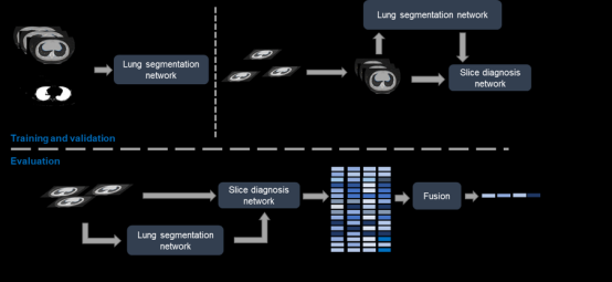
AI identification and diagnostic system for COVID-19
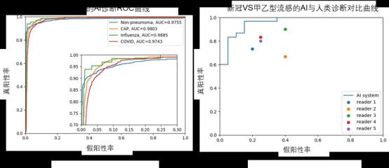
AI diagnostics versus expert diagnostics on a large-scale test set of COVID-19
In this study, the feasibility of using CT to accurately distinguish between COVID-19 and influenza A/B pneumonia is increased, and the AI+CT scheme is expected to become one of the distinguishment schemes. In a diagnostic comparative experiment with five frontline doctors, the AI system generally outperformed the five doctors in distinguishing COVID-19 from the other two types of pneumoniae. During the flu season in winter when the outbreaks of influenza A and B can occur again, accurate diagnosis is important for the prevention and control of both COVID-19 and influenza A/B pneumonia.
With the developed AI diagnostic tools, the team performed a subgroup analysis on large datasets. With the analysis of the diagnostic performance of different population (age, gender, etc.), the team found that age has a great influence on the diagnostic performance, and speculated that this conclusion could be attributed to the fact that the elderly population tends to exhibit a wider range of pulmonary lesions. The team systematically compared the diagnostic properties of X-ray and CT, and verified the performance advantage of CT over X-ray. They also found that the results of X-ray were more accurate when CT motion artifacts were larger. This provides a reference for optimizing the imaging program of patients with pneumonia.
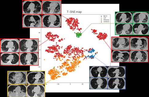
Spatial distribution of characteristics of three types of pneumonia samples and non-pneumonia samples
The paper was published in Nature Communications in October 2020. Jin Cheng (2012 Ph.D. student from the Department of Automation, whose supervisor is Professor Zhou Jie), Chen Weixiang (2017 Ph.D. student from the Department of Automation, whose supervisor is Professor Zhou Jie), and Cao Yukun (Wuhan Union Hospital of China) are the co-first authors of the paper, and Associate Professor Feng Jianjiang and Professor Shi Heshui are the co-corresponding authors. This paper was supported by the Special Fund of Zhejiang University for Covid-19 Prevention and Control.
Jin, C., Chen, W., Cao, Y. et al. Development and evaluation of an artificial intelligence system for COVID-19 diagnosis. Nature Communications 11, 5088 (2020).